Oral manifestations of systemic and discoid lupus erythematosus in a population of patients attending an Oral Medicine department: a retrospective study
OR4
Shir-Lynn Tan
Simone Liguori, Marco Carrozzo
Background:
Lupus erythematosus (LE) affects the mouth in 40% of patients, with oral ulcerations being most frequently reported. Oral LE can present, both clinically and histologically, as lesions resembling oral lichen planus (OLP), making differential diagnosis challenging. This retrospective study aims to characterise oral LE in a group of patients attending the Newcastle Dental Hospital Oral Medicine Department, assessing any possible correlation between oral lesions and patients’ clinical characteristics.
Methodology:
The clinical course of 13 patients with LE was evaluated. Diagnosis was established according to the 2019 EULAR/ACR classification criteria and histopathological examination of oral tissue samples were reviewed.
Results:
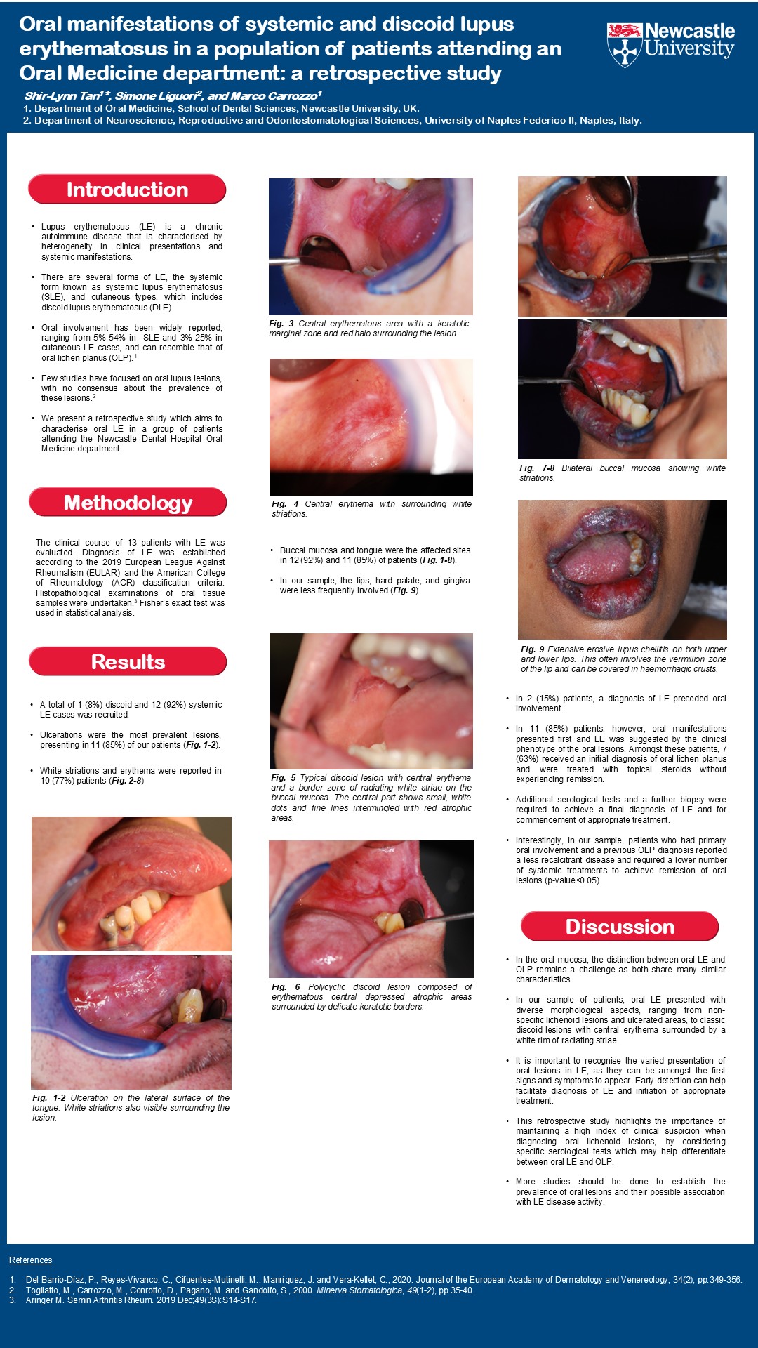
A total of 1 (8%) discoid and 12 (92%) systemic LE cases was recruited. Ulcerations were the most prevalent lesions (85% of patients), while white striations and erythema were reported in 77% of patients. Buccal mucosa and tongue were the affected sites in 12 (92%) and 11 (85%) patients, while the lips, hard palate and gingiva were less frequently involved. In 2 (15%) patients, a diagnosis of LE preceded oral involvement. In 11 (85%) patients, however, oral manifestations presented first and LE was suggested by the clinical phenotype of the oral lesions. Amongst these patients, 7 (63%) received an initial diagnosis of OLP and were treated with topical steroids without experiencing remission. Additional serological tests and a further biopsy were thus required to achieve a final diagnosis of LE and for commencement of appropriate treatment. Interestingly, patients who had primary oral involvement and a previous OLP diagnosis reported a less recalcitrant disease and required a lower number of systemic treatments to achieve remission of oral manifestations (p-value<0.05).
Conclusion:
This retrospective study highlights the importance of maintaining a high index of clinical suspicion when diagnosing oral lichenoid lesions by considering specific serological tests to help differentiate between oral LE and OLP.

