Raised lesion on the hard palate- what tumour could this be?
CR10
Manpreet Kaur
Manpreet Kaur, Daniel Brierly, Muzzammil Nusrath
Introduction:
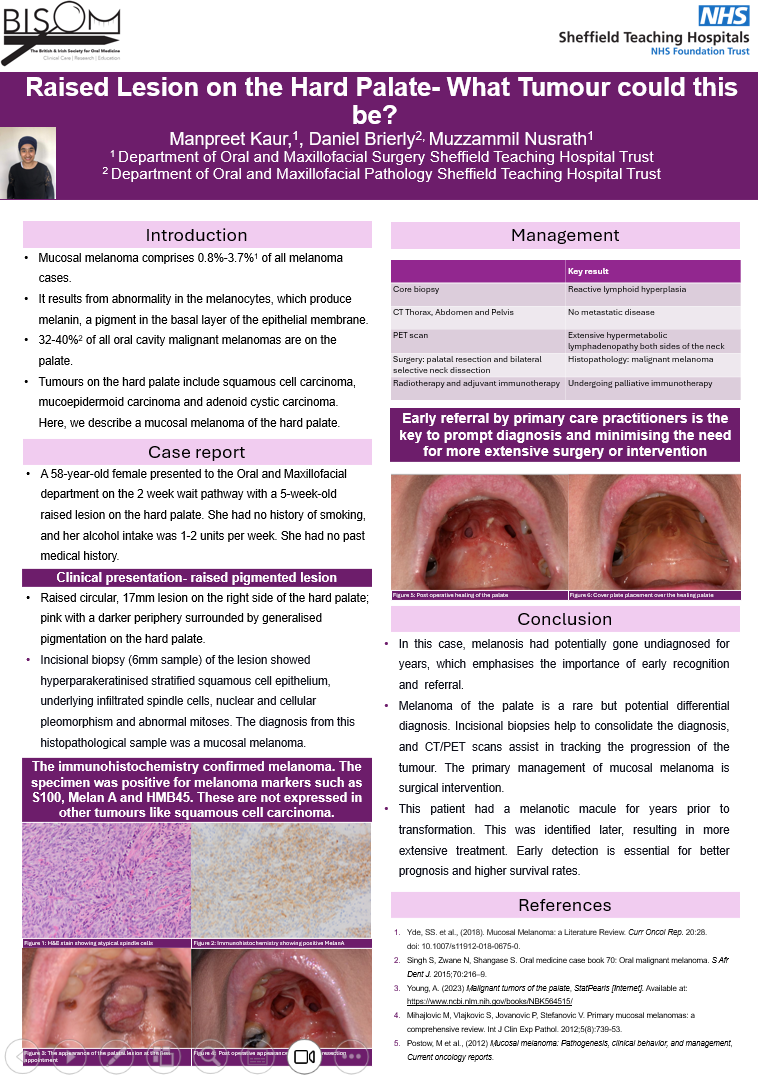
Mucosal melanoma comprises 0.8%-3.7% of all melanoma cases. It results from abnormality in the melanocytes, which produce melanin, a pigment in the basal layer of the epithelial membrane. 32-40% of all oral cavity malignant melanomas are on the palate. Tumours on the hard palate include squamous cell carcinoma, mucoepidermoid carcinoma and adenoid cystic carcinoma. Here, we describe a mucosal melanoma which developed on the hard palate.
Case Report:
A 58-year-old Female presented following a two-week wait referral from the General Dental Practitioner to the Oral and Maxillofacial Outpatient Department with a 5-week-old, raised 1cm lesion on the hard palate. This patient then had an incisional biopsy of the lesion, which showed hyperparakeratinised stratified squamous cell epithelium, underlying infiltrated spindle cells, nuclear and cellular pleomorphism and abnormal mitoses. The diagnosis from this histopathological sample was a mucosal melanoma. A core biopsy showed reactive lymphoid hyperplasia. After further staging scans, she had surgery (bilateral selective neck dissection and palatal resection). After this surgical procedure, she had radiotherapy and adjuvant immunotherapy, as her neck nodes identified malignant melanoma. This patient had a melanotic macule for years before transformation but was not identified, which resulted in more extensive treatment.
Conclusion:
In this case, she potentially had undiagnosed melanosis for years, which emphasises the importance of early referral. Melanoma in the palate is rare but is a potential differential diagnosis. Incisional biopsies help to consolidate the diagnosis, and CT/PET scans assist in tracking the progression of the tumour. The primary management of mucosal melanoma is surgical intervention. Early detection is essential for better prognosis and higher survival rates.

